| 📌 The essentials The MASAI (Mammography Screening with Artificial Intelligence) trial, published in The Lancet on January 31, 2026, is the largest randomized controlled trial of AI in any cancer screening program ever conducted. In 105,934 women across Sweden, AI-supported mammography improved screening sensitivity from 73.8% to 80.5% (p=0.031) while specificity remained identical at 98.5% in both groups (p=0.88). The interval cancer rate, the gold standard measure of missed cancers between screenings, was lower in the AI group: 1.55 versus 1.76 per 1,000 women screened. AI reduced aggressive and advanced interval cancers specifically, including fewer non-luminal A (more aggressive) tumors in the AI group (43 versus 59). And AI triaged 44% of scans to single-reader review without loss of accuracy, directly addressing radiologist workforce constraints. This post covers what the trial measured, how the AI worked, what the numbers mean in practice, and what remains open. |
|---|
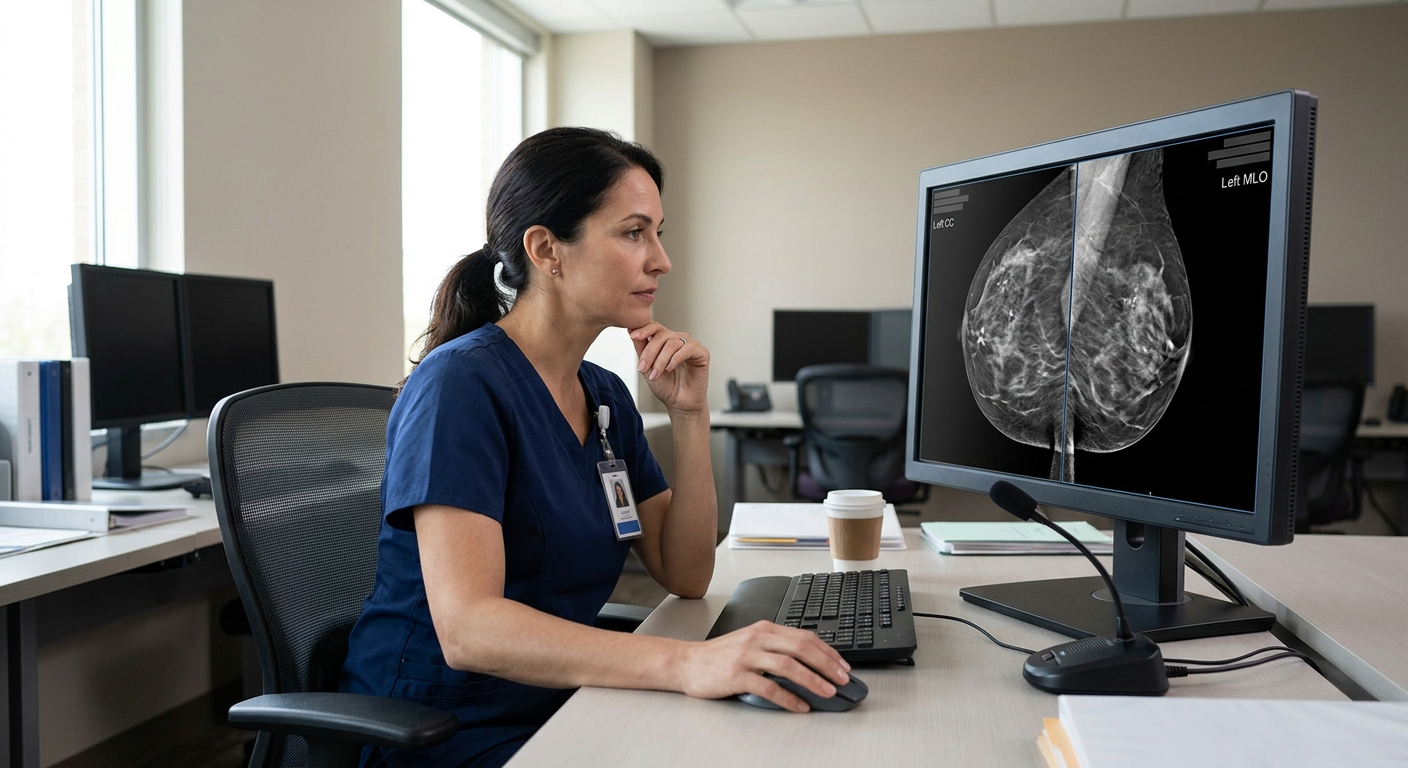
Every year in the United States, roughly 40 million mammograms are performed. Each one is read by at least one radiologist, and in many countries including Sweden, by two. Reading is time-consuming, cognitively demanding, and subject to the same variation in judgment that affects every human visual task. Radiologists miss some cancers. They also flag some findings as suspicious that turn out to be benign, sending women back for additional imaging or biopsies they did not need.
The promise of artificial intelligence in mammography is that it could do better on at least one of those problems without making the other worse. Catch more cancers while generating no more unnecessary callbacks. Or reduce the reading burden on an overstretched radiologist workforce while maintaining safety. Ideally, both.
The MASAI trial, published in The Lancet on January 31, 2026, is the first and largest randomized controlled trial of AI in any cancer screening program. It enrolled over 105,000 women in Sweden and ran from April 2021 to December 2022. The full results answer the central questions directly: AI-supported mammography caught more cancers and produced no increase in false positives.
The Measure That Matters Most: What Is an Interval Cancer?
Before getting into the numbers, it helps to understand what the MASAI trial was primarily designed to measure. The primary endpoint was not detection rate during screening. It was the interval cancer rate.
An interval cancer is a breast cancer diagnosed between scheduled screening rounds, meaning after a mammogram that came back negative. These are the cancers the screening missed. A woman left the screening appointment with a clean bill of health and developed a symptomatic cancer before her next scheduled appointment. Interval cancers tend to be more aggressive than screen-detected cancers because aggressive tumors grow faster and are more likely to become apparent between screening rounds rather than at the next scheduled scan.
Reducing the interval cancer rate is the gold standard test of whether a screening program improvement is real. It means the test is catching more of the dangerous cancers before they become symptomatic, not just generating more detections of indolent findings that would never have harmed the patient.
The MASAI Trial: Design and What AI Was Actually Doing
The MASAI (Mammography Screening with Artificial Intelligence) trial (NCT04666026) was a randomized, controlled, single-blinded, population-based screening accuracy trial conducted across three regions in Sweden. Enrollment ran from April 2021 through December 2022. A total of 105,934 women were randomly assigned, with 105,915 eligible for the final analysis: 53,043 in the AI-supported group and 52,872 in the standard double-reading group.
The median age in both groups was approximately 54 years, consistent with a population-based screening program. Sweden screens eligible women every 1.5 to 2 years, or annually for those at higher risk.
How the AI worked in this trial
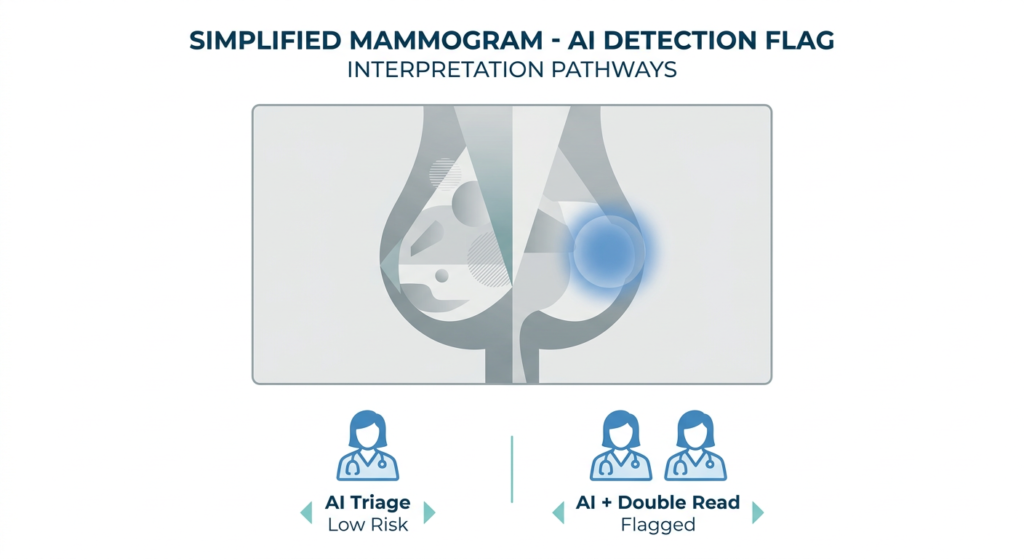
The AI system played two roles in the intervention arm. First, it triaged each mammogram scan for single or double reading by radiologists. Scans the AI assessed as lower risk were forwarded to a single radiologist read rather than the standard two-reader process. Scans assessed as higher risk received double reading with AI detection support. Second, in double-read cases, the AI highlighted suspicious areas on the images to assist the radiologists reviewing the scan.
The AI system used in MASAI was trained, validated, and tested on over 200,000 mammography scans before deployment. The control arm received standard double reading by two radiologists without any AI involvement.
The Results: What the Trial Found
| Outcome | AI-supported | Standard double-read |
|---|---|---|
| Sensitivity | 80.5% (95% CI 76.4 to 84.2%) | 73.8% (95% CI 68.9 to 78.3%) |
| p-value for sensitivity | p=0.031 | Reference |
| Specificity | 98.5% (95% CI 98.4 to 98.6%) | 98.5% (95% CI 98.4 to 98.6%) |
| p-value for specificity | p=0.88 (no difference) | Reference |
| Interval cancer rate (per 1,000) | 1.55 (95% CI 1.23 to 1.92) | 1.76 (95% CI 1.42 to 2.15) |
| Invasive interval cancers | 75 | 89 |
| T2+ stage interval cancers | 38 | 48 |
| Non-luminal A interval cancers | 43 | 59 |
| Reduction in radiologist workload | 44% of scans routed to single-read | All scans double-read |
The specificity finding is the critical reassurance
Sensitivity is the ability to detect cancer when it is present. Specificity is the ability to correctly clear patients who do not have cancer. The two are often in tension: systems designed to catch more cancers tend to generate more false alarms. The MASAI finding that specificity was identical at 98.5% in both groups (p=0.88) is therefore one of the most important numbers in the entire dataset. AI caught more cancers without generating more unnecessary callbacks or biopsies. That is the combination the field has been working toward.
What the interval cancer characteristics tell us
The numbers behind the 12% reduction in interval cancers are worth examining carefully. Women in the AI-supported group had fewer interval cancers that were invasive (75 versus 89), fewer that had reached T2 or larger size (38 versus 48), and fewer that were non-luminal A subtype (43 versus 59). Non-luminal A tumors are the more aggressive breast cancer subtypes, including triple-negative and HER2-positive cancers. Their reduction is particularly meaningful because these are the cancers where early detection makes the biggest difference to survival.
The lead author of the MASAI trial, Dr. Kristina Lang of Lund University’s Division of Diagnostic Radiology, noted in the published report that the trial found AI-supported screening improves the early detection of clinically relevant breast cancers, reducing aggressive and advanced cancers diagnosed in between screenings. She also noted at the time of publication that AI adoption must be done carefully, with tested tools and continuous monitoring.
A Second 2026 Study in Nature Cancer: AI Increased Detection From 7.54 to 9.33 Per 1,000 Women
The MASAI results are part of a broader pattern of evidence building in 2026. A separate study published in Nature Cancer reported that AI-supported mammography increased cancer detection from 7.54 to 9.33 per 1,000 women screened. That translates to roughly 1.8 additional cancers detected per 1,000 women in a given screening round, or about 1 in 556 women screened gaining a detection they would have missed under standard reading.
The two studies use different endpoints and populations, so direct numerical comparison is limited. Together, they strengthen the evidence that AI-supported mammography reading improves cancer yield in real-world screening settings, not just in retrospective analyses of selected image archives.
What This Means for Patients Who Get Mammograms Today
Is AI reading my mammogram now?
Possibly. Several FDA-cleared AI systems for mammography assistance are in use at imaging centers and hospitals across the United States, including Transpara (ScreenPoint Medical) and iCAD. The specific AI tool used in the MASAI trial is not the only one commercially available, and the evidence base for individual products varies. The MASAI trial result tells us that when a well-validated AI system is integrated into a structured screening workflow, the combined result outperforms standard double reading. It does not automatically apply to every AI product on every platform.
Does AI replace the radiologist?
No. In the MASAI trial design, AI triaged scans to single or double reading by radiologists and highlighted suspicious areas for radiologist review. A radiologist made every final read. The AI reduced how many scans required two radiologists’ time and provided detection support to the reader who reviewed each case. The result was a 44% reduction in the portion of radiologist reading time devoted to double reads, without loss of accuracy.
This matters for healthcare systems facing radiologist workforce shortages. The United States and many European countries have a well-documented shortage of breast imaging specialists. A technology that allows the same number of radiologists to safely read more scans without reducing quality addresses a real structural problem in cancer screening infrastructure.
Will AI increase false alarms?
The MASAI trial specifically answers this. Specificity was 98.5% in both groups and the difference was not statistically significant (p=0.88). This is a reassurance, not a trivial finding. An AI system that drove up the recall rate would expose women to unnecessary imaging anxiety and follow-up procedures. Maintaining specificity while improving sensitivity is the combination that makes AI integration clinically viable rather than just mathematically impressive.
| What interval cancers found in the study tell us about AI and aggressive tumors The 12% reduction in interval cancers in the AI arm is the most clinically meaningful finding for patients who actually get mammograms. Interval cancers are the ones that grow between screenings and become symptomatic before the next scheduled appointment. They tend to be more aggressive precisely because aggressive tumors grow faster. The MASAI data specifically showed the AI arm had fewer T2-or-larger interval cancers (38 versus 48) and fewer non-luminal A tumors (43 versus 59). Non-luminal A cancers are the harder-to-treat subtypes, including triple-negative and HER2-positive disease. Reducing the interval rate for these subtypes, not just for all cancers in aggregate, is what the trial’s authors describe as clinically relevant improvement. The benefit of higher sensitivity was consistent across age groups and breast density categories. Women with dense breast tissue, who are often told that mammography is less reliable for them, saw the same relative benefit from AI support as women with non-dense tissue. |
|---|
What the Study Does Not Tell Us
The MASAI results are strong and the trial design is rigorous. Honest presentation of the evidence also requires naming what remains open.
This trial used one specific AI system. The results apply to the validated tool used in MASAI. There are multiple AI mammography products on the market with varying levels of clinical evidence behind them. FDA clearance for a device does not automatically mean its performance matches the MASAI AI system in this structured workflow.
Long-term survival data is not yet reported. The trial measured interval cancer rates and tumor characteristics, not survival outcomes. Whether the improved early detection translates into reduced breast cancer mortality over 10 to 20 years is the most important unanswered question. Based on what we know about how interval cancer rates relate to mortality in breast screening, the expectation is that it does, but long-term data from this cohort will be needed to confirm.
The trial was conducted in Sweden. Sweden has a national, population-based screening program with standardized protocols. Results may differ in healthcare systems with more fragmented screening delivery, different population characteristics, or different baseline double-reading rates.
Not all AI reads improve on human performance equally. A secondary analysis of the trial noted that the sensitivity improvement applied to invasive cancers but not to in-situ cancers specifically. Understanding which cancer types AI improves detection for, and which it does not, matters for interpreting the clinical impact.
Practical Guidance for People Due for a Mammogram
- If you are due for a mammogram and have been putting it off, this study does not change the recommendation to screen. It strengthens it. Current American Cancer Society guidelines recommend annual mammograms starting at age 40 for average-risk women.
- If your imaging center uses AI-assisted reading, it is reasonable to ask which system they use and whether it has been prospectively validated in clinical trials, not just retrospective analyses.
- If you receive a callback for additional imaging after a mammogram, that is not necessarily a sign something went wrong. Recall rates remained the same under AI-supported reading in this trial. Most callbacks do not result in a cancer diagnosis.
- For women with dense breast tissue who have been told mammography is less sensitive for them: the MASAI data showed the AI benefit was consistent across breast density categories. That is an encouraging finding, though supplemental screening options remain a separate conversation to have with your provider.
- Screening intervals have not changed based on this evidence. The MASAI results strengthen the case for regular mammography participation, not for altering how often you screen.
For related women’s health coverage on Health Evidence Digest, see our post on new 2026 cervical cancer screening guidelines that now allow self-collection for HPV testing, as well as our coverage of pembrolizumab becoming the first approved immunotherapy for ovarian cancer.
Sources
MASAI trial registration: NCT04666026. ClinicalTrials.gov.
ASCO Post coverage: Randomized Trial Shows AI-Supported Mammography Improves Sensitivity and Lowers Interval Cancer Rate. The ASCO Post. February 2, 2026.
EurekAlert/Lancet press release: AI-supported mammography screening results in fewer aggressive and advanced breast cancers, finds full results from first randomized controlled trial. EurekAlert. January 29, 2026.
AJMC coverage: AI-Supported Mammography Caught More Cancers During Screening. AJMC. 2026.
Lund University press release: AI support in breast cancer screening: Fewer missed cancer cases. Lund University. January 30, 2026.
MASAI interim safety results (2023): Lång K et al. Artificial intelligence-supported screen reading versus standard double reading in the Mammography Screening with Artificial Intelligence trial (MASAI): a clinical safety analysis of a randomised, controlled, non-inferiority, single-blinded, screening accuracy study. The Lancet Oncology. 2023;24(8):936-944.
MASAI AI detection analysis (2024): Lång K et al. Identifying normal mammograms in a large screening population using artificial intelligence. Lancet Digital Health. 2024. doi:10.1016/S2589-7500(24)00267-X
Patient resources: American Cancer Society mammography guidelines | National Cancer Institute | Dense Breast Info
| Disclaimer: Health Evidence Digest provides general information about health research for educational purposes. This content does not constitute medical advice and is not a substitute for consultation with a qualified healthcare provider. Mammography screening recommendations should be discussed with your physician based on your individual health history and risk factors. |
|---|
| Disclaimer: Health Evidence Digest provides general information about health research for educational purposes. This content does not constitute medical advice and is not a substitute for consultation with a qualified healthcare provider. Mammography screening recommendations should be discussed with your physician based on your individual health history and risk factors. |